Secrets of how cells cram in oversized genomes revealed

Secrets of how cells cram in oversized genomes revealed
Stanford researchers have shown how the goopy material inside bacterial cells and interactions with other biomolecules encourage DNA segments to fold up to a thousandth of their actual length.
A new study reveals key details about how bacterial cells manage to pack in their chromosomal DNA a thousand times longer than the cells themselves, and — even more amazingly — do so in a highly organized manner. The research, led by Stanford biologists, offers new insights into the fundamental yet poorly understood process whereby genomes fold up in a way that allows specially tailored access by certain biomolecules.
"How cells package and compact their oversized genomes in a way that is not random is a huge question in biology," said Christine Jacobs-Wagner, the Dennis Cunningham Professor in the School of Humanities and Sciences and professor in biology. Jacobs-Wagner began this study with her lab at Yale and completed the research at Stanford when she moved to Stanford Humanities and Sciences in 2019. "With this study, we've taken a step forward in answering this question."
The study examined why the DNA segments of chromosomes in a bacterial cell preferentially interact with themselves rather than the surrounding, goopy contents of the cell, known as cytoplasm. This interaction causes the DNA to compact down into a mesh-like mass — dubbed a nucleoid — that is full of holes. These holes have to be in the right places and of a certain size, however, to allow for biomolecules such as ribosomes to access messenger RNAs made from genes to convert them into proteins. The nucleoid, therefore, cannot simply be a balled-up, tangled mess.
The nucleoid avoids becoming a jumble and instead attains organization, the study suggests, in part because of repulsive interactions between DNA and RNA. Presumably because of their respective chemistries and size, DNA and RNA basically "try to get away from each other" said Jacobs-Wagner. This apparent repulsive interaction causes DNA to spread out in areas where the genes that make RNA are active and RNA is abundant, and scrunch up in areas where RNA is scarce. As a result, in sections of DNA where the genes that make RNA are particularly active, the nucleoid has a bigger mesh size, conveniently making it easier for ribosomes to access and assemble on the RNAs emerging from these active DNA regions. In contrast, in DNA regions where genes are not transcribed frequently, the nucleoid is dense, making these active regions less accessible.
Overall, this delicate interplay between biomolecules forms a super-squished yet structured nucleoid.
"This study just goes to show that there is still a lot about cells we don't know," said Jacobs-Wagner, the study’s senior author and an institute scholar at Stanford ChEM-H. "How a chromosome gets folded inside a cell in such a precise, organized manner is really fundamental to life but is not something we fully understand."
A fresh approach
The study, published online July 8 in the journal Cell, innovatively took a page from polymer physics, a field which studies large molecules built up from smaller, repeating subunits — like DNA, for instance. In polymer physics, researchers often examine what goes on chemically in a mixture of two substances, such as when polymers are immersed in solvents (usually liquids).
Taking that tack, the study's researchers modeled how DNA as a polymer would behave when interacting with the cytoplasm as a solvent. Polymer physicists broadly classify solvents as good, poor, or ideal based on how the polymer and solvent interact. In a "good" solvent, the polymer preferentially interacts with the solvent, whereas in a "poor" solvent, the polymer prefers to interact with itself, while something in between is an ideal solvent.
"If a polymer 'likes' a solvent, the polymer will stretch in this good solvent to maximize interaction," explained Jacobs-Wagner. "But if a polymer doesn't like the solvent, that is, finds this poor solvent to be repulsive, the polymer will instead come together and condense."
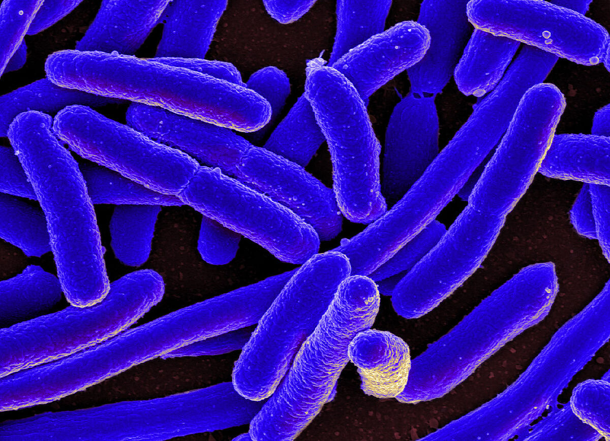
For their polymer physics-based model of DNA and cytoplasm, the researchers selected the well-studied bacterium Escherichia coli (E. coli). By estimating the possible mesh (hole) size of the nucleoid and the concentrations of DNA in the nucleoid, the researchers were able to show that the cytoplasm effectively acts as a poor solvent, thus encouraging the DNA segments to crumple up into a nucleoid.
To learn more about how this crumpling occurs and leads to nucleoid organization, Yingjie Xiang experimented with living E. coli. Xiang was a graduate student in the Jacobs-Wagner lab at Yale before the Jacobs-Wagner lab moved to Stanford in 2019. One technique they used, called fluorescence microscopy, involved using dyes and tagging certain biomolecules with a glowing protein to observe the density of DNA and other biomolecules in different regions of a nucleoid. The researchers also delved deeply with a technique called cryo-electron tomography by collaborating with Professor Jun Liu and his postdoc Yunjie Chang at Yale University. It begins with freezing a biological sample, then slicing the sample into thin sections, and finally imaging the sections with an electron microscope.
Examining E. coli with these techniques revealed that DNA was less dense in areas where ribosomes were clumped together, and DNA was more compact in regions where ribosomes were scarce. Furthermore, experimentally blocking transcription (the process of converting DNA into RNA) and translation (the process of ribosomes “reading” RNA to make proteins), results in more compact, denser DNA. These results suggest that the acts of transcription and translation actively shape the nucleoid, while contributing to the cytoplasm acting as a poor solvent.
Looking into other cells
While the study focused on unraveling genome folding in E. coli, Jacobs-Wagner said that the approach her lab developed is generalizable to other kinds of single-celled bacteria, and multicellular eukaryotes, such as humans, who have more complex cell types. As with bacteria, just how multicellular creatures package and organize their genomes still needs more research in order to understand genome folding.
In eukaryotic cells, the task of DNA folding is greatly aided by proteins called histones. DNA winds around these proteins, just like thread wraps around a spool. Bacteria lack histones and have had to evolve different strategies to compact their DNA.
However it is done, genome folding is an impressive display of natural engineering, orderly compacting a roughly 1.5-millimeter-long genome into an E. coli cell measuring just a micrometer (millionth of a meter) long. In the case of humans, about two meters of DNA must coil down over a 100,000-fold to nestle within a 10 micrometer-wide nucleus.
"There is much we still have to learn about genome folding in all three domains of life, namely bacteria, eukaryotes like ourselves, and in the single-celled organisms called archaea" said Jacobs-Wagner. "It really is amazing how cells do this."
About the School of Humanities and Sciences
The School of Humanities and Sciences is the foundation of a liberal arts education at Stanford. The school encompasses 23 departments and 25 interdisciplinary programs across the arts, humanities, natural sciences, and social sciences. It is the university's home for applied and fundamental research, where free, open, and critical inquiry is pursued across disciplines.


