High-resolution look inside living cells now possible without fluorescence
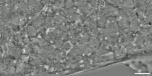
A microscope developed at Stanford can reveal the interaction of nanostructures inside living cells at the highest resolution yet, about 120 nanometers, without the use of fluorescent labels. The technology could lead to a host of new advances in the life sciences.
In brief:
- The new microscope blends two imaging approaches to reveal tiny parts of living cells and allow nearly unlimited observation time, without labels.
- The method provides greater context, allowing researchers to view movement of organelles and other cellular structures as well as their responses to pathogens and drugs.
- This new way of seeing into cells could lead to breakthroughs in a range of fields. Stanford researchers are already using it to study the uptake of cancer drugs into cells and plant–microbe interactions.
The view into living cells just got better.
Stanford researchers have combined two microscopy techniques to create a one-of-a-kind instrument that can show cell structures interacting in real time at an unprecedented 120-nanometer resolution—the highest achieved without the use of fluorescent labels.
This new “label-free” technology, called Interferometric Image Scanning Microscopy, or iISM, will allow scientists to observe cellular structures in their wider context, including their responses to intrusions, such as pathogens or drugs. The advance is detailed in the Nature portfolio journal Light: Science and Applications.
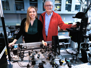
“This new microscope provides a fantastic new view into the cell, where you can see the tiny structures and machines in the cell moving, changing, and interacting without having to add fluorescence to observe them,” said senior author W.E. Moerner, the Harry S. Mosher Professor of Chemistry in Stanford’s School of Humanities and Sciences. “It’s a wonderful look into these complex little cellular boxes that drive our life.”
The iISM’s capabilities could enable breakthroughs in a range of life science fields, from understanding disease mechanisms and developing drugs to investigating relationships between plants and microbes.
While the resolution is not as fine as other types of specialized microscopes, iISM’s label-free method has many advantages: It allows researchers to see many structures at once for long observation times. In contrast, methods that rely on fluorescence to label structures typically can highlight only a few target structures at a time. The fluorescence can eventually “bleach” or wear off. These labels can also be difficult to introduce and, in some cases, may change the behavior of the structures they tag.
The iISM can also operate at a substantially lower illumination power than comparable high-contrast label-free approaches, reducing the risk of photodamage in living cells and making it less likely to disturb the tiny, delicate structures being observed.
This does not mean that the new microscope will replace the use of fluorescence microscopy, which has yielded insights in the life sciences for decades, said first author Michelle Kueppers, a postdoctoral scholar in Moerner’s lab.
“Every method has its advantages and disadvantages, and we believe in a complementary implementation in the future,” Kueppers said. “If we use the strengths of fluorescence for molecular specificity and the strength of iISM for label-free context and dynamics, we can really start tackling questions that have been difficult to address before.”
Many ‘eyes’ on the same point
The iISM manages to achieve higher resolution and sensitivity by combining the advantages of two different microscopy methods—a combination that speaks to the expertise of the two co-authors. Moerner, who won the 2014 Nobel Prize in chemistry for his work on super-resolution fluorescence microscopy, specifically sought to bring Kueppers to Stanford because of her doctoral work focused on “interferometric scattering microscopy.”
Scattering is what makes the sky look blue. When light hits small particles—as when sunlight passes through the atmosphere and encounters dust, water droplets, and other molecules—it deviates from its path and scatters in different directions. The particles in Earth’s atmosphere scatter the short waves of blue light more strongly than red light, so the sky appears blue to human eyes.
In the interferometric scattering microscope, a laser shines on a cell and the tiny structures inside scatter some of the light. A second laser beam is then used to amplify the weaker scattered light, making it strong enough to be detected so the small structures can be viewed.
The key advance of the iISM is the combination of the interferometric scattering method with an adapted concept from next-generation confocal microscopes. While traditional confocal microscopes use a pinhole and a single detector to focus on target structures, advanced versions of these microscopes use camera-based array detectors, which can capture many views of the same area.
For the iISM, the Stanford researchers used a type of array detector that enables the collection of more light than the pinhole and single detector model. This provides increased depth and precision. It works similar to the way two human eyes both take in information to distinguish the foreground from background—only the iISM uses not just two “eyes” but tens to hundreds of views from an array detector. The researchers then developed a method to combine these measurements into sharper, higher-contrast images.
The result is a label-free microscope capable of about 120 nanometer resolution while using less laser power and maintaining imaging speed—which means researchers can view living cells longer and more gently.
Wide vision for wide applications
Moerner and Kueppers are now working to further improve this technology and make it widely available to other scientists.
They have already started three collaborations with other Stanford researchers. One uses the new microscope to see the interaction among plant cells, fungi, and bacteria in real time. Another collaboration uses the iISM to see how a cancer drug is taken up by a cell, and a third planned effort will investigate how red blood cells change shape when encountering a malaria infection.
“This is not a niche technique,” Kueppers said. “It has broad applications, and we hope the life science community will be well served by it, leading to many new discoveries.”
Acknowledgments
This research received support from the U.S. National Institute of General Medical Sciences.
Media contact:
Sara Zaske, School of Humanities and Sciences, 510-872 -0340, szaske [at] stanford [dot] edu (szaske[at]stanford[dot]edu)


Pollution linked to fish hybrids in Mexican river